
Endoscopic Lumbar Discectomy for Left L4-L5 Disc Herniation

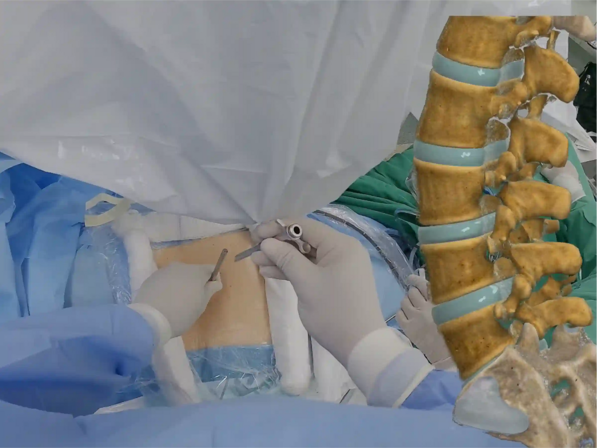
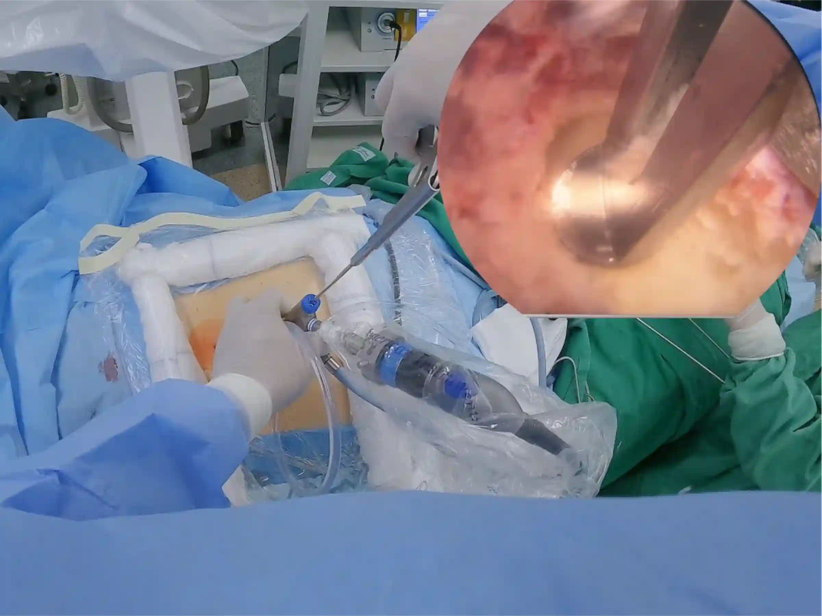
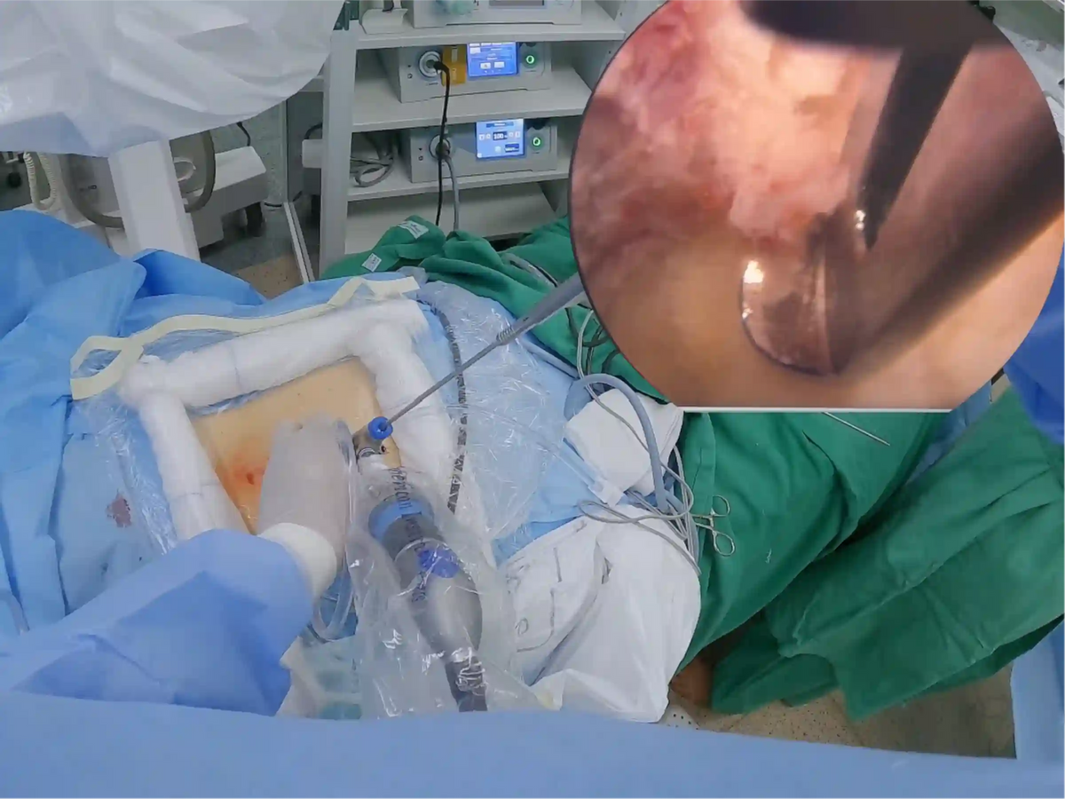
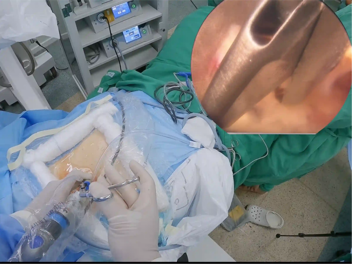
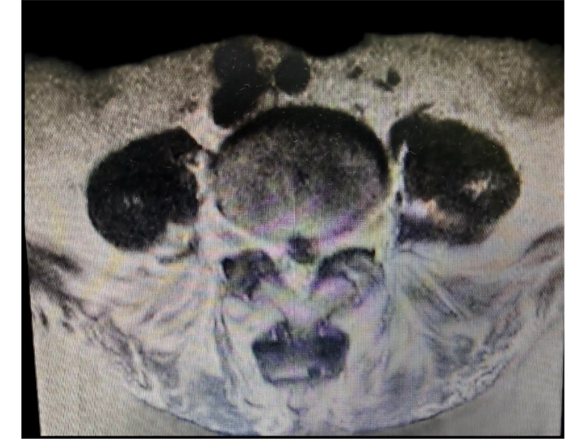
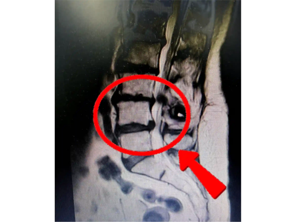
This educational case follows an endoscopic lumbar discectomy for a left L4-L5 disc herniation in the setting of an adjacent prior interspinous or interlaminar device at the level above. The teaching focus is on building orientation from the lamina-facet junction, confirming bony landmarks, exposing the superior and inferior laminae, defining the medial and lateral boundaries of the interlaminar window, and planning controlled enlargement of the window before opening the ligamentum flavum.
The case highlights procedural reasoning during endoscopic access, including fluoroscopic confirmation, use of the working cannula, soft-tissue clearance, endoscopic bipolar hemostasis, radiofrequency-assisted exposure, and transition from cutting burr to diamond burr as the surgeon approaches deeper cortical bone. It also demonstrates how Kerrison rongeur technique and instrument direction are discussed in relation to the underlying dural sac and neural structures.
Endoscopic spine surgery requires visual-spatial orientation because the surgeon relies heavily on endoscopic visualization and tissue appearance during dissection; working-channel endoscopes and longer endoscopic instruments differ from open instruments in length, angulation, depth markings, and tactile feedback (KB1, page 15). Endoscopes can provide magnification, illumination, depth visualization, and direct positioning over the operative field, while angled endoscopes introduce additional orientation and navigation considerations (KB3, page 14). The case also reinforces caution during powered bone removal, as an endoscopic drill can move with rotational force when contacting bone; maintaining controlled hand support is described in the literature as a way to reduce inadvertent movement risk (KB2, page 22).
This content is intended for medical education and procedural analysis only. It does not replace supervised surgical training, institutional protocols, clinical judgment, or formal review by qualified spine specialists.
INCLUDED CONTENT
- Case-based procedural narration translated and adapted into English for educational use.
- Stepwise review of left-sided endoscopic access to the L4-L5 level.
- Anatomical orientation around the lamina, facet, spinous process, and interlaminar window.
- Discussion of endoscopic soft-tissue exposure and hemostasis.
- Drilling strategy for interlaminar window enlargement.
- Kerrison rongeur technique considerations during thin cortical bone removal.
- Safety-oriented commentary on working near ligamentum flavum and neural elements.
- Knowledge-base supported notes on endoscopic visualization, working-channel instruments, and powered drill caution.
TARGET AUDIENCE
- Neurosurgery residents and fellows
- Spine surgery fellows
- Orthopedic spine trainees
- Surgeons learning endoscopic lumbar spine anatomy and procedural workflow
- Medical educators preparing spine endoscopy teaching material
- Operating room professionals seeking anatomy-focused procedural context
EDUCATIONAL NOTICE
For professional medical education only. This material does not provide medical advice, does not guarantee clinical outcomes, and is not a substitute for supervised surgical training, institutional protocols, or review by qualified medical professionals. Human expert review is required before educational publication or use.
Escolha opções
